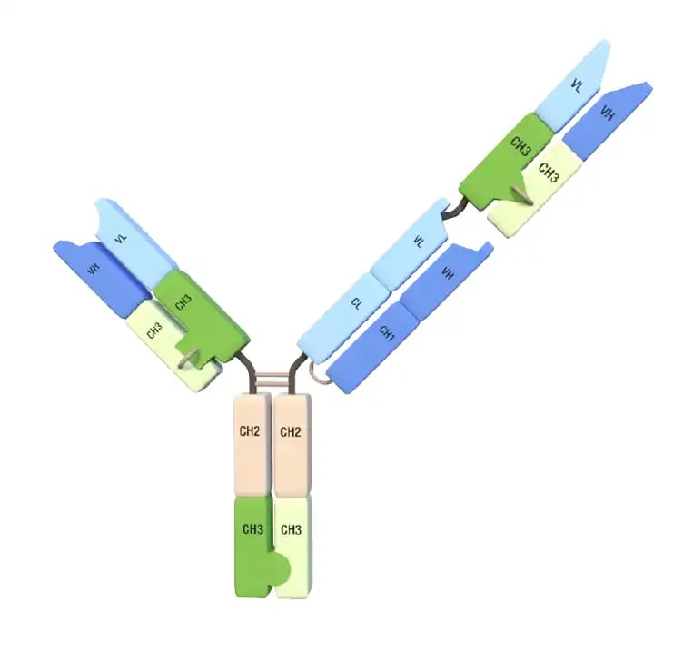
At a glance
-
Less than 1% of an injected ADC dose binds tumor cells, and normal-tissue expression of common targets caps the dose. A third binding arm addresses both limits rather than one at a time.
-
HER2 × HER2 × HER3 internalizes faster and to a greater extent in SK-BR-3 cells than trastuzumab, pertuzumab, or our own HER2 × HER3 bispecific. In JIMT-1 mice, two doses of the DXd conjugate prevented tumor growth across the study, while every comparator arm grew.
-
EGFR × VEGF × HER3 only binds and internalizes where VEGF is present. That raises killing in the EGFR-high line and leaves cells largely intact in the EGFR-low line when VEGF is absent, which is the safety window the design targets. In vivo studies are next.
-
Both molecules in Invenra’s T-Body™ platform feature mAb-like CMC: up to 1.5 g/L in transient CHO, concentration to 100 mg/mL, Protein A and IEX purification, and cysteine conjugation to a uniform DAR of 6 at 98.85% monomer.
Belinda Wenke, PhD, presented "From Bispecific to Trispecific Antibodies: Reimagining the Next Generation of ADCs" at PEGS Europe 2026.

The full recorded presentation is available on demand below. This guide is a written companion that covers the same material, including our latest trispecific ADC data.
Multispecific antibodies open up avenues in the ADC field that monoclonals alone have trouble accessing, or simply can’t.
Target two tumor antigens, and you work to close off antigen loss as an escape route. Target a matrix component alongside a tumor antigen, and you can reach tumors where the extracellular matrix blocks access. Add biparatopic binding or conditional activation, and you can work on safety and efficacy at the same time.
Here, we walk through the exposure problem conventional ADCs run into, the four mechanisms we've been building, and preclinical data on two programs: HER2 × HER2 × HER3, a biparatopic-plus trispecific, and EGFR × VEGF × HER3, which only binds and internalizes where VEGF is present.
The data is current as of publication. These programs are active, and results will change as studies read out.
Conventional ADCs deliver too little payload, and toxicity caps the dose
Less than 1% of an injected ADC dose binds to tumor cells, and even less enters them and releases the payload as designed.
Three things compound the problem:
First, delivery is restricted. Antigen heterogeneity and tumor stroma limit how much drug reaches the tumor, and stromal cells can physically occlude access.
Second, durability erodes. Alternative pathways come online during treatment, and cancer cells with low antigen density escape.
And third, the dose is capped. The payloads are potent and effective, but normal-tissue expression of many targets produces on-target, off-tumor toxicity that narrows the therapeutic window.
The result is insufficient tumor payload, short durability, and constrained dosing.
Why a third arm via a trispecific?
Our case for trispecific (TsAb) ADCs rests on four central points, each tied to the limits we just covered.
- More cancer indications covered. Targeting multiple targets increases the number of cancers likely to respond.
- Broader patient coverage within an indication. Targeting multiple targets increases the number of patients likely to respond within a given cancer.
- Better tumor control. Binding multiple targets limit the tumor's escape routes and attack a larger portion of a heterogeneous tumor. Covering escape mechanisms at the time of first treatment doesn't give those mechanisms time to develop, and when they do start to develop, the other arms can address them quickly.
- More mechanisms of action. The elaborated architecture of a trispecific enables mechanisms that a bispecific or a mAb can't access. Our first programs group under HYPER-Internalization™, meaning guided and enhanced internalization and so greater payload delivery into the cell, and we get there two ways: biparatopic binding and a VEGF-driven approach we call the VEGF Activated Avidity Lock, or VAAL. (Targeting three tumor antigens is the third while stromal plus tumor targeting is the fourth.)
Four suits, four mechanisms
To organize the formats and target combinations into something you can hold in your head, we sort them into a deck of cards. Each suit carries a different mechanism of action.
These are example cards from the platform deck:
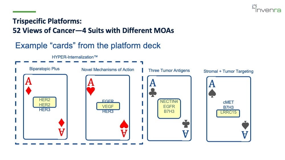
Four example playing cards for trispecific ADC designs: Ace of diamonds, biparatopic plus, HER2 x HER2 x HER3. Ace of hearts, novel mechanisms of action, EGFR x VEGF x HER3. Ace of clubs, three tumor antigens, NECTIN4 x EGFR xB7H3. Ace of spades, stromal plus tumor targeting, cMET x B7H3 x LRRC15. The diamonds and hearts suits of the card deck are grouped under HYPER-Internalization.
These are example cards from our platform deck:
|
Suit |
Mechanism |
Example targets |
|
Diamonds |
Biparatopic plus |
HER2 × HER2 × HER3 |
|
Hearts |
Novel mechanisms of action, such as VAAL |
EGFR × VEGF × HER3 |
|
Clubs |
Three tumor antigens |
NECTIN4 × EGFR × B7H3 |
|
Spades |
Stromal plus tumor targeting |
cMET × B7H3 × LRRC15 |
Diamonds and hearts both sit under HYPER-Internalization™, and they are one area where a trispecific gives an advantage over its mAb and bispecific counterparts. Those two are the focus of the rest of this article.
The card framing extends to 52 Views of Cancer, our deck of playing cards illustrated by cancer patients, survivors, and family members.
The B-Body bispecific and T-Body trispecific platforms
Invenra arose from generating bispecifics and building stable platforms to explore that modality.
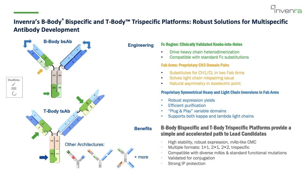
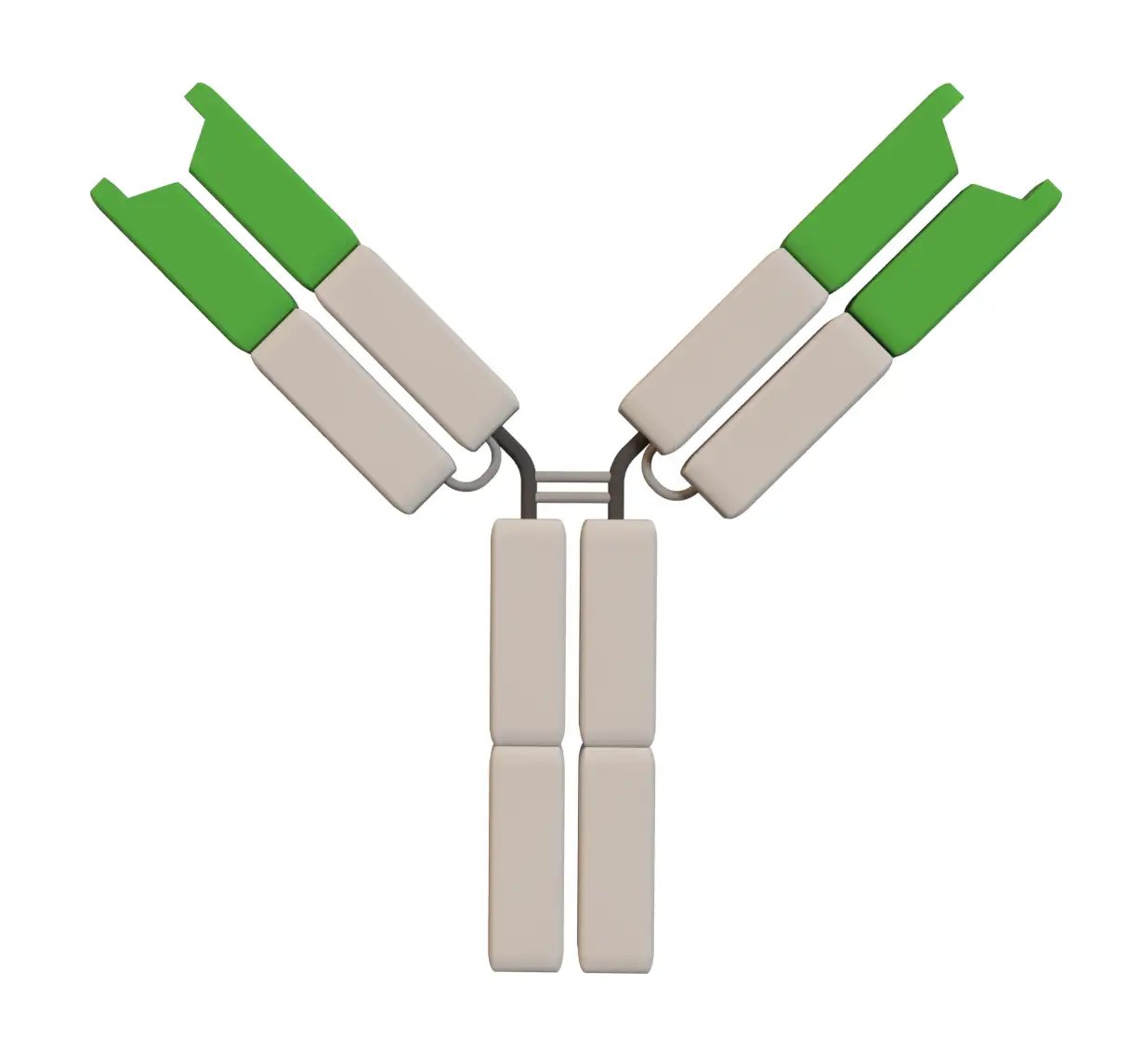
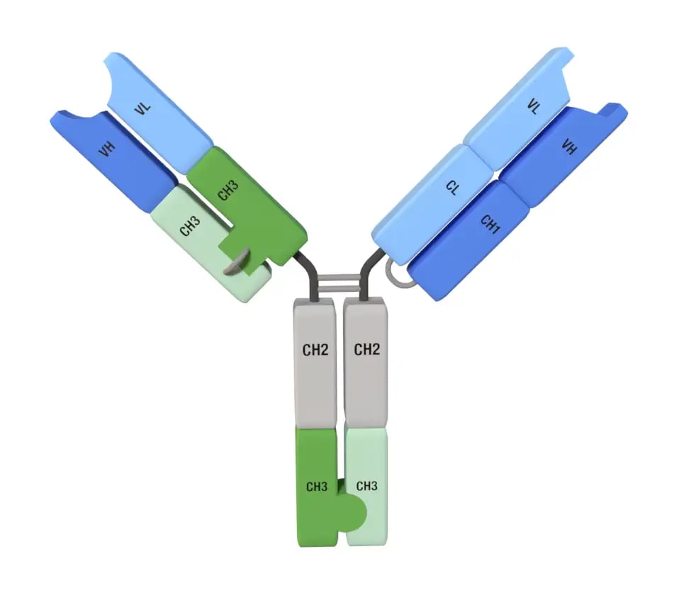
Diagrams of a B-Body bispecific and a T-Body trispecific antibody, with the knobs-into-holes Fc, the proprietary CH3 domain pairs in the Fab arms, and the symmetrical heavy and light chain inversions labeled, alongside other supported architectures.
The B-Body® bispecific platform rests on two solutions:
- For heavy chain pairing, we use clinically validated knobs-into-holes, which drive heterodimerization. That helps on the manufacturing and purification side, and on the biology side, it leaves room for the standard Fc substitutions used to tune effector function and PK.
- For light chain pairing, we use proprietary engineered CH3 domain pairs that substitute for CH1/CL in one to two Fab arms. These domains support both kappa- and lambda-derived antibodies, so monoclonal antibodies can be converted to a bispecific format without compromising stability or creating purification challenges. They also introduce natural asymmetry in the isoelectric point of the arms, which makes purification fast and robust, and makes screening fast and robust as well. That matters in practice as it lets us screen many bispecific combinations and pick the best molecules out of a given set of monoclonals.
We also use proprietary symmetrical inversions of the heavy and light chains in the Fab arms, which maintain expression yields, keep purification efficient, and support the plug-and-play screening we rely on to find the best arms to combine.

The T-Body™ trispecific platform is part of the same lineage. It builds on the bispecific using another orthogonal engineered CH3 domain pair.
The bispecific domain and the trispecific domain are completely orthogonal: they don't interact with each other, and they don't interact with the Fc. The pairing is high fidelity, and the molecules don't come apart during purification.
That robustness is what makes it practical to explore the architectures available to trispecifics, including adding Fab arms above or below the Fc, depending on the function you want.
Together, the two platforms give a simple, accelerated path to lead candidates, with high stability, robust expression, and mAb-like CMC; multiple formats including 1×1, 2×1, 2×2, and trispecific; compatibility with diverse mAbs and standard functional mutations; validation for conjugation; and strong IP protection.
HER2 × HER2 × HER3: biparatopic plus
Why these three specific arms?
The design comes out of the properties of the targets and how they interact with each other.
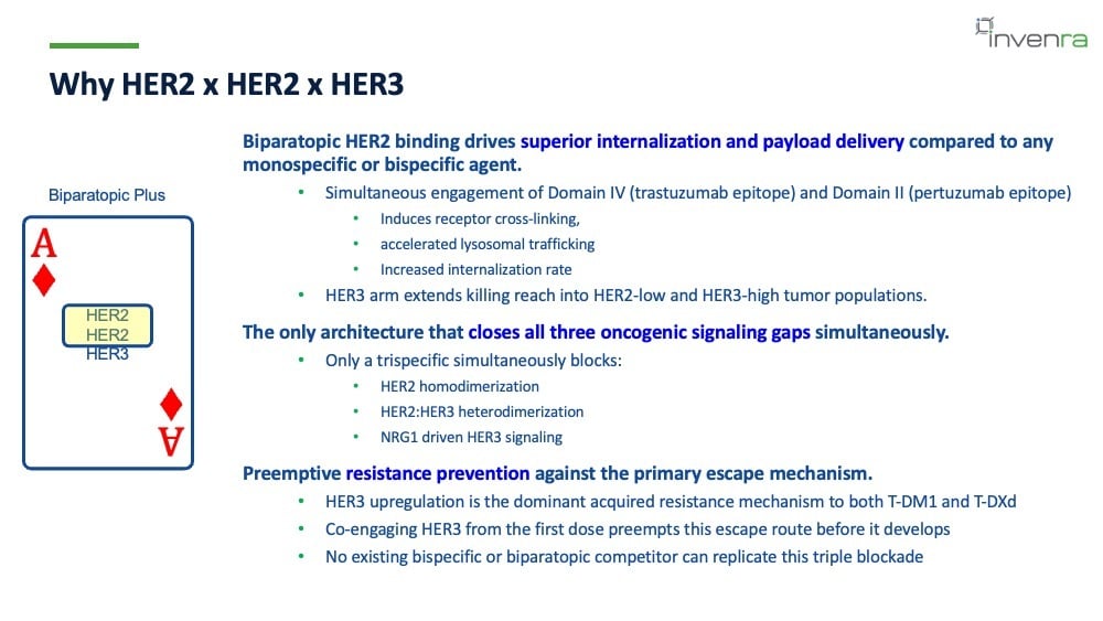
HER2 already has effective therapies on the market, and the literature shows that internalization can be the limiting step in many cases. Engaging the trastuzumab epitope on Domain IV and the pertuzumab epitope on Domain II at the same time drives internalization: it induces receptor cross-linking, accelerates lysosomal trafficking, raises the internalization rate, and leads to enhanced killing.
The HER3 arm extends killing reach into HER2-low and HER3-high tumor populations.
The ERBB family receptors signal through each other and form heterodimers. The biparatopic HER2 binding blocks HER2 homodimerization. The HER3 arm blocks HER2:HER3 heterodimerization, and its epitope blocks NRG1 binding, thereby suppressing HER3 signaling from that direction as well.
The molecule targets all three oncogenic signaling routes simultaneously.
Resistance is the other half of the rationale. HER3 upregulation is a primary acquired resistance mechanism in HER2-driven cancers during exposure to trastuzumab, T-DM1, and T-DXd. Engaging HER2 and HER3 from the first dose is meant to block that escape route before it develops, and doing it simultaneously is something only a trispecific has been shown to do.
There's a question behind all of this, since a HER2 biparatopic and a HER2 × HER3 bispecific both already exist.
-
The biparatopic gets better internalization.
-
The HER2 × HER3 bispecific helps prevent escape.
Each has its own potential liabilities. What we wanted to know was whether two approaches that are already somewhat validated could be combined into one molecule and give the best of both.
Let's go a little deeper into our performance data for this trispecific.
Developability and ADC compatibility
On the T-Body platform, HER2 × HER2 × HER3 shows:
- IgG-like CHO transient yields up to 1.5 g/L
- Stable CHO pools in progress at 1+ g/L, from unoptimized growth conditions
- Concentration up to 100 mg/mL, which puts subcutaneous dosing in range
- Greater than 95% purity by SEC
- Protein A and IEX compatibility
- A half-life of 8.8 days in hFcRn Tg32 mice
Disulfide conjugation is validated, with a maximum DAR of 6 for the standard trivalent T-Body trispecific. The ceiling is structural: the disulfides in the B-Body and T-Body domains are buried and inaccessible.
Conjugated with MC-GGFG-DXd, the molecule runs at 98.85% monomer by SEC with an LC-MS DAR of 6, at a good retention time.
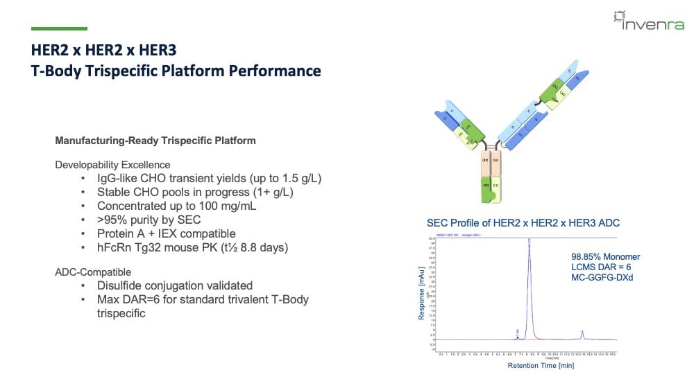
Developability and ADC compatibility results for HER2 × HER2 × HER3, with a T-Body schematic and the SEC profile of the conjugated ADC showing 98.85% monomer, LC-MS DAR of 6, and MC-GGFG-DXd.
Stable CHO cell line development is in progress, and the results so far are consistent. Two stable pools, after a single-step Protein A purification, gave this:
|
Stable pool |
Protein A titer |
Purified |
Main peak by SEC |
|
P3 |
1 g/L |
0.83 g/L |
74% |
|
P4 |
1 g/L |
0.76 g/L |
75% |
On CE-SDS, the trispecific is the dominant band in both pools, with very few impurities. (These are pools, not final clones, and we expect a meaningful bump in both yield and purity when we move to clones. That's been our experience with the bispecifics.)
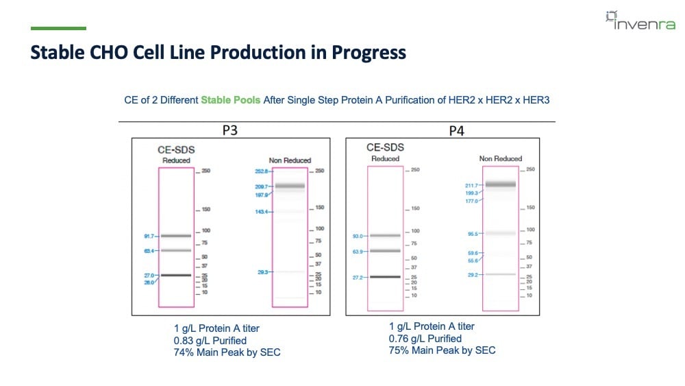
Reduced and non-reduced CE-SDS profiles of two stable CHO pools, P3 and P4, of HER2 × HER2 × HER3 after single-step Protein A purification.
Formulation for our bispecific programs has been simple and hasn't needed exotic excipients, and the trispecifics behave the same way.
In 10 mM sodium acetate, pH 5.2:
|
Formulation |
Tm1 (°C) |
Tagg (°C) |
Diffusion interaction parameter (mL/g) |
|
No excipients |
63.0 |
74.4 |
21.67 |
|
+ 3% proline |
63.0 |
64.2 |
30.54 |
|
+ 4.5% sorbitol |
63.9 |
73.0 |
15.01 |
|
+ 2.9% sucrose |
63.0 |
73.1 |
17.33 |
Melting and aggregation temperatures shift slightly between formulations without much change in stability as measured by percent monomer, and the molecule withstands several freeze/thaw cycles without significant degradation.
Across five cycles, concentration held steady in all four formulations and percent monomer stayed between roughly 96% and 99%, with the largest decline in the formulation with no excipients.
The diffusion interaction parameter is worth reading closely here. Negative values indicate self-association. Here every formulation, including the one with no excipients at all, gives large positive numbers, which signal very low self-association.
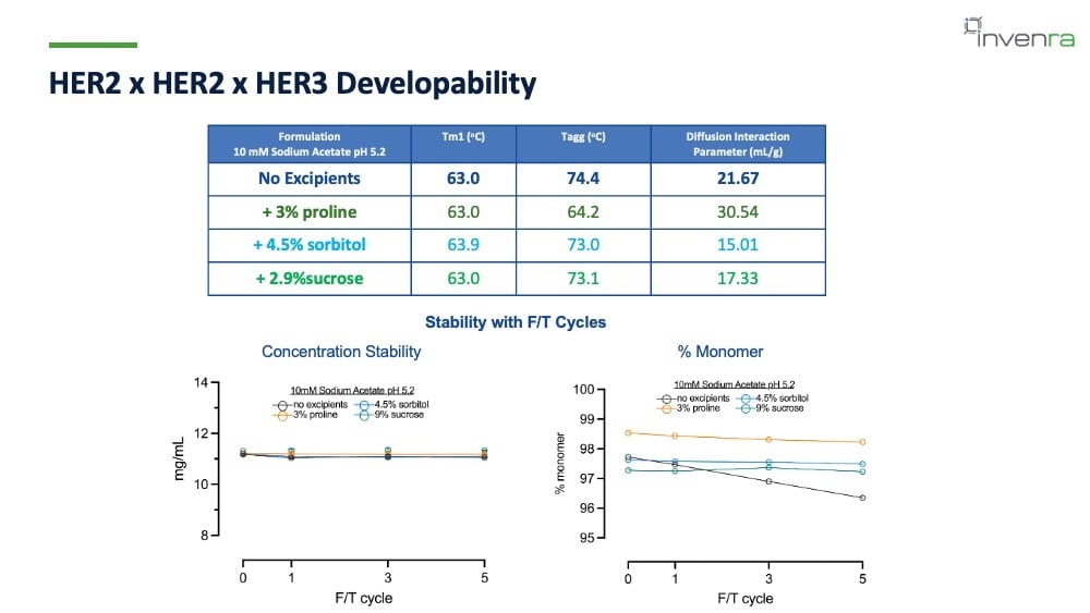
Table of Tm1, aggregation temperature, and diffusion interaction parameter for four formulations of HER2 × HER2 × HER3, with graphs of concentration and percent monomer across five freeze/thaw cycles.
Internalization
In SK-BR-3 breast cancer cells measured over 12 hours, the trispecific antibody achieves a higher total amount internalized than the comparators and does so at a faster rate.
The comparison runs against our own HER2 × HER3 bispecific, trastuzumab, and pertuzumab. Trastuzumab alone is the lowest curve.
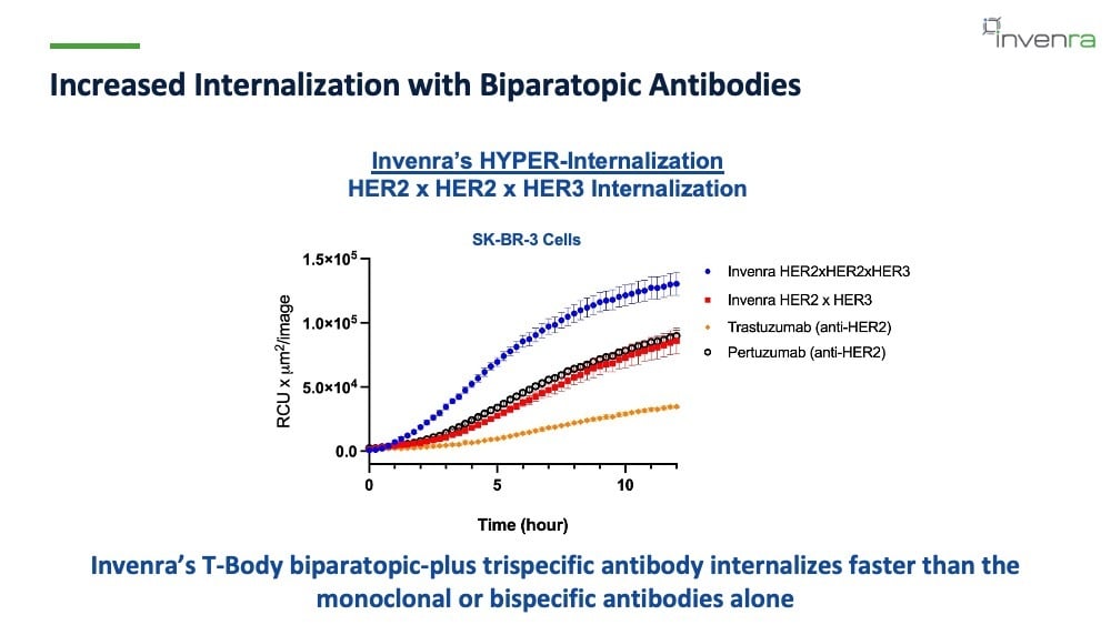
Internalization over 12 hours in SK-BR-3 cells, measured as RCU times square microns per image. HER2 × HER2 × HER3 rises highest and fastest, followed by pertuzumab and the HER2 × HER3 bispecific, with trastuzumab lowest.
In vitro ADC killing
These are piggyback assays (an anti-Fc Fab conjugated to MMAE rather than our intended payload). This is a benchmarking experiment.
The toxin is held constant across the tool molecules and the cell lines, so what varies is the antibody and the target copy number, and the cell lines are chosen to vary copy number rather than to represent the indication.
|
Cell line |
HER2 copies |
HER3 copies |
|
SK-BR-3 |
250,000 |
9,500 |
|
NUGC-4 |
54,000 |
24,000 |
|
T-47D |
18,000 |
13,300 |
The comparators are zanidatamab (a HER2 biparatopic), patritumab (a HER3 mAb), trastuzumab (a HER2 mAb), and a negative control.
Where HER2 is very high, in SK-BR-3, the trispecific curve sits with the HER2 comparators rather than clearly ahead of them. The separation shows up as expression drops. In NUGC-4 and T-47D the trispecific is left-shifted against the mAbs and the biparatopic and reaches a lower percent viability.
That pattern is one of the things we've learned building these. As molecules get more complex, the assay and the cell line matter more, and you have to be deliberate about which question each assay is asking. Running several cell lines across a range of expression levels is how the trispecific's contribution becomes visible at all.

Piggyback ADC killing curves with Fab MMAE in SK-BR-3, NUGC-4, and T-47D cells, comparing HER2 × HER2 × HER3 with zanidatamab, patritumab, trastuzumab, and a negative control, with HER2 and HER3 surface copy numbers below each panel.
In vivo efficacy
The xenograft study used JIMT-1, a trastuzumab-resistant breast cancer line, in 11-week-old female NOD SCID mice.
Cells went in subcutaneously at 5 × 10⁶ on day -7. Mice received two intraperitoneal doses, on day 0 once tumors had grown out and again on day 11, with molar equivalent dosing normalized to a 5 mg/kg dose for the control IgG1.
The conjugates were made by cysteine conjugation or purchased as biosimilars. Zanidatamab-DXd is a tool molecule, built the same way as the other conjugates for the sake of comparison, which is why the DARs differ across arms.
|
Group |
Treatment |
DAR |
|
1 |
DPBS |
n/a |
|
2 |
HER2 × HER2 × HER3-DXd |
6 |
|
3 |
Zanidatamab-DXd |
4 |
|
4 |
Trastuzumab-DXd |
8 |
|
5 |
Patritumab-DXd |
8 |
|
6 |
Control hIgG1-DXd |
8 |
Tumors in the trispecific ADC group showed no growth across the study. The other arms all rebounded. Zanidatamab-DXd held tumors suppressed until around day 30, then started to grow. Trastuzumab-DXd grew roughly linearly. Patritumab-DXd grew fairly quickly.
The DPBS and control IgG1 groups grew out right away, reaching about 1,700 mm³ by the end of the study. Per-animal curves for each group are on the second in vivo slide, with day 0 tumor volumes between 126 and 142 mm³ across groups.
Two caveats here: This was our first look at in vivo efficacy with a trispecific ADC, and the study has no HER2 × HER3 bispecific ADC arm, which would have been a useful control.
The question we were asking was narrower than "is this the best ADC":
With the payload standardized across arms, can we see the antibody itself providing the benefit?
The data shows we can. Whether more can be pulled out of this molecule by optimizing the linker and payload is a separate question, and an open one!
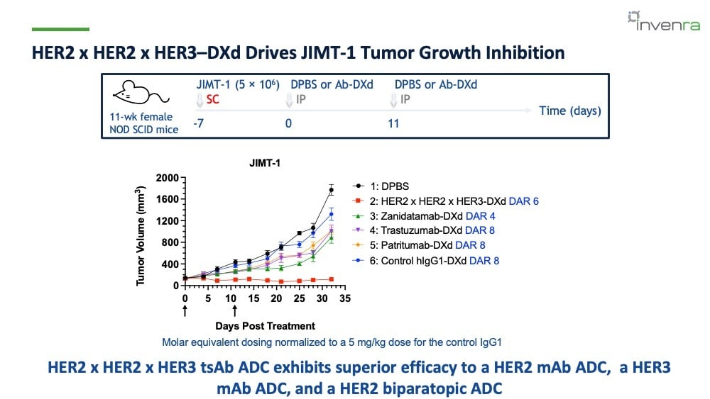
Study design and mean tumor growth curves in JIMT-1 NOD SCID mice. Tumor volume in the HER2 × HER2 × HER3-DXd group stays flat near baseline through day 32 while all other groups grow.
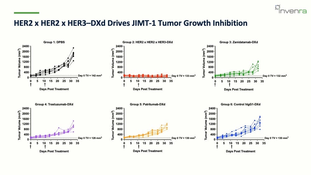
Per-animal tumor growth curves for all six groups.
EGFR × VEGF × HER3: the novel-mechanism
Again, why these three specific arms?
These targets were chosen primarily because of on-target, off-tumor toxicity. EGFR is notorious for skin toxicity. EGFR, and to some extent HER3, are expressed on normal skin, in the GI tract, and in the kidney, which leaves high-affinity monospecific ADCs inherently dose-limited.
The resistance argument is the same shape as the HER2 one. HER3 upregulation is the primary resistance axis in EGFR-driven cancers, so this target combination addresses escape routes that are clinically known to emerge after treatment.
We're not the only ones who think the pair is worth going after. Izalontamab brengitecan (BL-B01D1), a first-in-class EGFR × HER3 bispecific ADC with FDA Breakthrough Therapy Designation, reported a 66% objective response rate in TKI-pretreated EGFR-mutant NSCLC at WCLC 2025. What the VAAL arm adds on top of a validated pair is conditional tumor selectivity.
How the VEGF Activated Avidity Lock works
VEGF is an obligate homodimer. It's present in the tumor microenvironment where it drives signaling and vascularization. We expect its concentration to be higher there than in normal tissue, with constant cycling keeping that difference in place.
Because VEGF is a homodimer, every VEGF dimer presents two epitopes for the VEGF arm to bind. When VEGF is present and two antibodies are bound to the same dimer, the arms that aren't targeting VEGF are held at very high local concentration and can bind two targets on a cell avidly.
Take the VEGF away and the tumor-targeting arm is on its own. It's deliberately low affinity, so the antibody comes on and comes off with nothing tethering it to the cell, and it simply falls off. No significant engagement, no internalization.
Put the VEGF back and the picture changes: the VEGF arms bind, avid binding to the tumor antigen is supported, and internalization follows from that proximity. The therapeutic window widens because the molecule only engages cells where VEGF is around.
In our slide's words, it converts the tumor's own hypervascularization into an ADC activation switch.
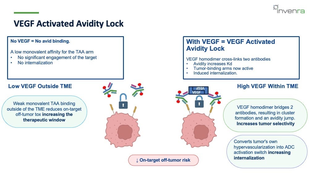
Diagram of the VEGF Activated Avidity Lock. Outside the tumor microenvironment, where VEGF is low, antibodies bind weakly and fall off. Inside it, where VEGF is high, a VEGF homodimer bridges two antibodies, producing cluster formation, an avidity jump, and internalization.
The avidity effect in other VEGF-binding molecules
The same effect shows up in other molecules that bind VEGF.
With Akeso's ivonescimab, a PD-1/VEGF bispecific, PD-1 is loaded on the sensor and VEGF is added during the dissociation phase; the dissociation rate slows. These are strong binders, so the effect is subtle, but the slope of the dissociation phase changes.
With CStone's CS2009, a PD-1/VEGF/CTLA-4 molecule, and with ivonescimab in a PD-1 reporter assay, the EC50 shifts lower in the presence of VEGF, indicating more potent PD-1 blockade.
Our own EGFR/VEGF molecule, T6690, behaves the same way: as VEGF concentration increases, the dissociation of EGFR flattens out, and the dissociation rate slows.
One caveat when reading these experiments is that they're looking at roughly a 1:1 interaction between molecules and VEGF, so the avidity effect becomes apparent at about a 1:1 ratio, though it's still present at lower ratios.
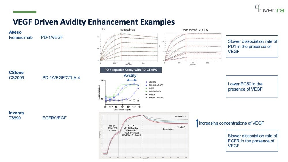
Binding traces for Invenra's EGFR/VEGF molecule T6690, showing slower EGFR dissociation as VEGF concentration increases.
Triggered binding with the EGFR × VEGF bispecific
We tested the mechanism first in a bispecific.
A549 tumor cells expressing EGFR at about 52,000 copies per cell were incubated with EGFR × VEGF bispecific antibodies with or without VEGF for six hours, then fixed, permeabilized, and stained with a secondary antibody, so fluorescence marks where antibody is.
Without VEGF, there's essentially one glowing spot in the field. With VEGF, the cells are decorated with the bispecific, and there's a punctate signal inside the cells where antibody has accumulated. The quantified green fluorescence tells the same story: signal only in the EGFR × VEGF plus VEGF condition, with a VEGF mAb control, a cells-plus-secondary control, and a cells-only control all flat.
In piggyback ADC assays using an anti-human-Fc Fab conjugated to MMAE, run in A549 (EGFR about 52,000 copies) and NCI-H1975 (EGFR about 32,000 copies), the killing curves shift left in the presence of VEGF in both lines.
The differential between the plus-VEGF and minus-VEGF conditions is large.
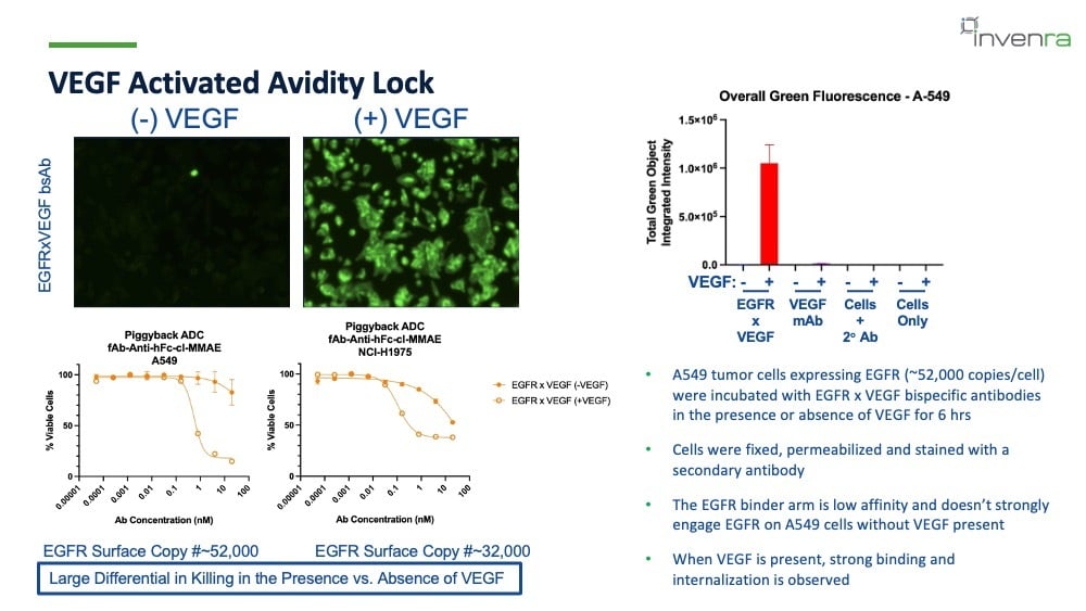
Fluorescence images of A549 cells treated with an EGFR × VEGF bispecific without and with VEGF, a bar chart of total green fluorescence by condition, and piggyback ADC killing curves in A549 and NCI-H1975 cells with and without VEGF.
Triggered internalization with the trispecific
Moving to the trispecific adds a second tumor target to control. We ran internalization over 12 hours in two cell lines with different copy numbers, against cetuximab (an EGFR mAb) and izalontamab (an EGFR × HER3 bispecific):
|
Cell line |
EGFR copies |
HER3 copies |
|
A549 |
52,000 |
<1,000 |
|
NUGC4 |
14,000 |
22,000 |
In both lines, the trispecific internalizes only when VEGF is present. Without VEGF, the curve is flat. The comparators stay flat in both conditions, which is what you'd expect given that neither has a VEGF-interacting arm.
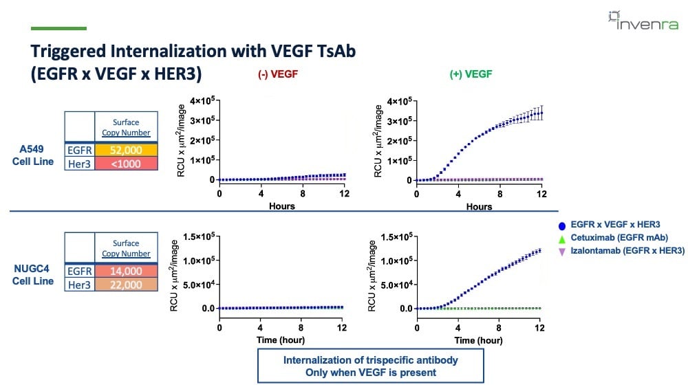
Internalization over 12 hours in A549 and NUGC4 cells, minus and plus VEGF. The EGFR × VEGF × HER3 trispecific rises only in the plus-VEGF panels; cetuximab and izalontamab stay at baseline throughout.
The FabFluor assay reads out the same thing in a different way. The dye fluoresces at low pH, so the signal appears once antibody has been internalized and degraded in the lysosome.
In A549 cells at eight hours, the trispecific shows very little signal without VEGF and a large increase with it. Cetuximab and izalontamab show almost nothing in either condition.
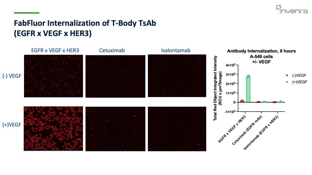
FabFluor images and a bar chart of antibody internalization in A549 cells at eight hours, with and without VEGF, for the EGFR × VEGF × HER3 trispecific, cetuximab, and izalontamab.
Killing and the safety window
The piggyback killing assay used 4 nM antibody plus MMAE Fab over five days, in the same two lines, against cetuximab, patritumab, and izalontamab.
In A549, the comparators bring viability down only modestly, and adding VEGF changes them little. The trispecific is the molecule that moves the needle: viability drops sharply once VEGF is present. The VEGF arm increases killing.
NUGC4 shows the other half of the mechanism. EGFR is lower here, which is closer to what background tissue looks like. The comparators kill potently whether or not VEGF is there. The trispecific doesn't.
Without VEGF, it leaves most cells viable, and with VEGF, it kills about as potently as the others. That difference is the safety window.
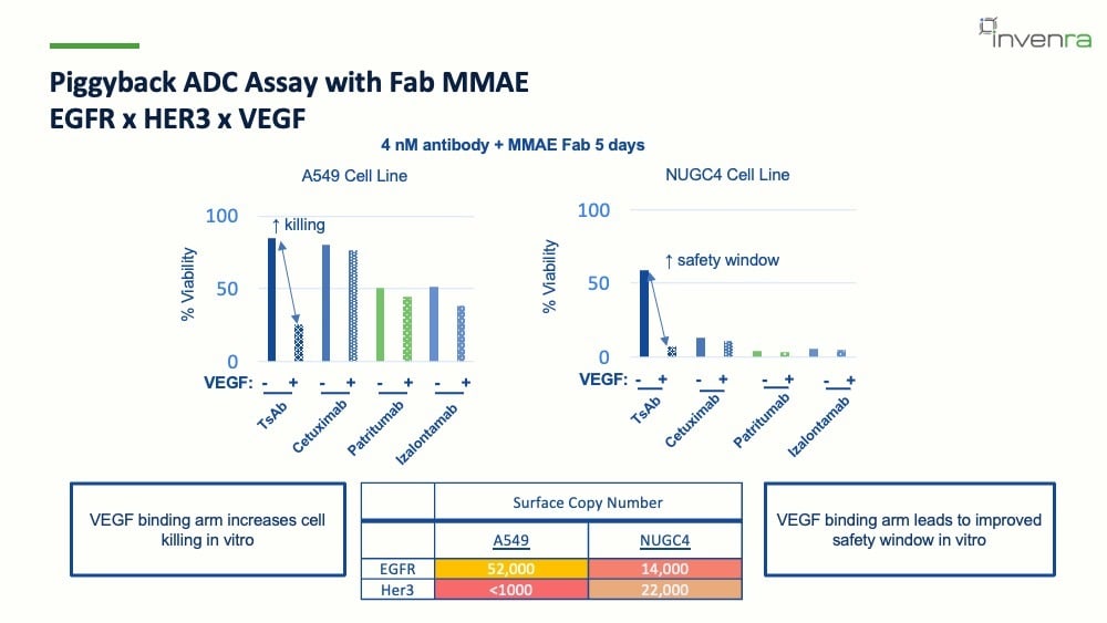
Bar charts of percent viability in A549 and NUGC4 cells after five days with 4 nM antibody plus MMAE Fab, minus and plus VEGF, for the trispecific, cetuximab, patritumab, and izalontamab, with EGFR and HER3 surface copy numbers below.
The advantages of a trispecific
Advancing next-generation ADCs means working on three interconnected challenges at once: a narrow therapeutic window, resistance and heterogeneity, and chemistry and manufacturing complexity.
Trispecific ADCs can address several of the issues that limit traditional mAb ADCs:
- Resistance and heterogeneity, by targeting multiple tumor-associated antigens simultaneously
- A poor normal-to-tumor expression window, by using mechanisms like VAAL to make avid binding conditional
- Poor internalization, since biparatopic-plus antibodies and the VEGF lock both increase it
- Dense stroma limiting access to the tumor, by targeting stroma as well as tumor through an "or" gate
Of course, none of that matters if the molecules can't be made. The reason we achieve these results is that these trispecifics can be manufactured and screened robustly enough to find the ones that work.
Where these two programs stand
HER2 × HER2 × HER3:
- Transient CHO expression at 1.5 g/L Protein A titer, stable CHO pool in progress at 1 g/L
- Concentration up to 100 mg/mL, freeze/thaw stability, and low self-association by diffusion interaction parameter
- Half-life of 8.8 days in hFcRn Tg32 mice
- Improved internalization and in vitro killing over HER2 or HER3 mAbs in piggyback ADC assays
- Proven cysteine conjugation to a uniform DAR of 6
- In vivo efficacy in JIMT-1 exceeding trastuzumab-DXd, patritumab-DXd, and zanidatamab-DXd
EGFR × VEGF × HER3
- Triggered internalization demonstrated in vitro
- Improved safety window in in vitro assays
- Next step: in vivo efficacy
Working with Invenra
Our B-Body® bispecific and T-Body™ trispecific platforms are built for rapid generation of developable multispecific constructs with expression yields up to 11 g/L for bispecifics, CMC-ready formats, subcutaneous compatibility, and standard downstream processing.
Our portfolio includes more than 30 active programs, with two antibody constructs advancing to clinical development through partner programs.

We work three ways across monoclonals, bispecifics, and trispecifics: custom discovery from target to lead, rapid prototyping from validated antibodies or partner mAbs, and licensable assets. Characterization sits alongside all of it, so the antibody engineers who built your molecule are the ones who characterize it.
One contract, one team, one set of materials.
If you're developing an ADC and want to work out whether a third arm helps with your targets and your tumor setting, get in touch and start the conversation.
Talk to us
If you're developing an ADC and want to work out whether a third arm helps with your targets and your tumor setting, get in touch and start the conversation.
 White paper
The Plug-and-Play Advantage of Invenra’s Multispecific Platforms
Invenra’s B-Body® and T-Body™ modular multispecific antibody platforms enable plug-and-play assembly and format-agnostic purification to deliver consistently high-purity bispecific and trispecific antibodies.